

Many patients still need a real Swan-Ganz alternative.
Now there is one.
Minimally invasive Right Heart and Venous Return Monitoring, when "non-invasive" is just not enough.
Currently not commercially available!

SVC Flow Pattern
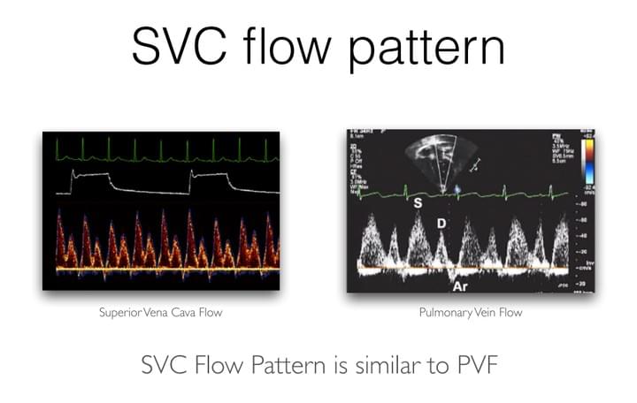
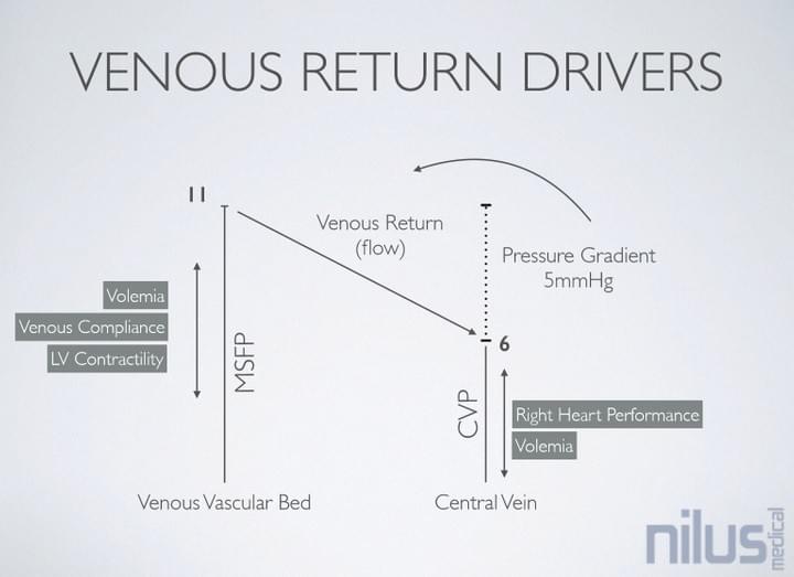
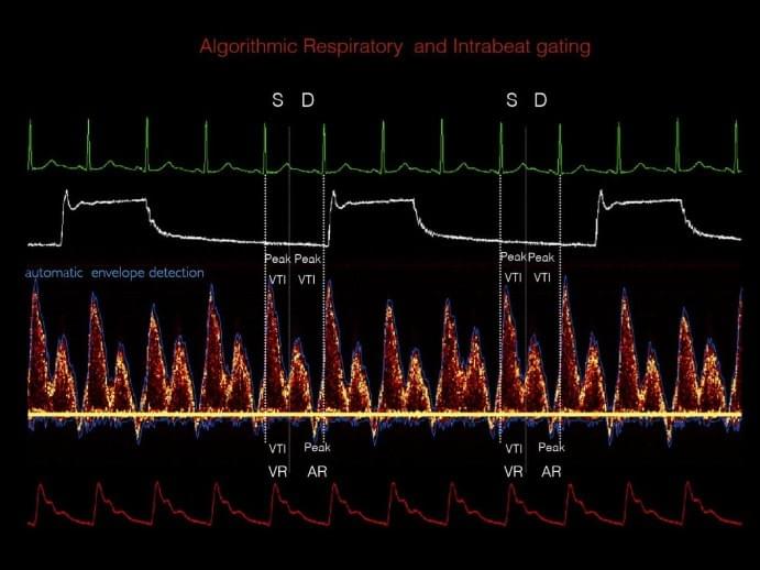
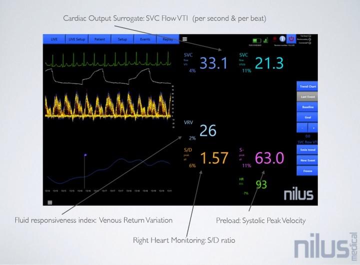
Superior Vena Cava blood flow pattern correlates to Right Heart function, Preload, and Fluid Responsiveness.
Familiar sonography parameters with easy to obtain signal via regular central line. No need to calibrate. Real-time, right-sided hemodynamic monitoring.

Continuous ultrasound monitoring helps to detect and discern changes early
With Nilus, the Superior Vena Cava flow monitoring provides the missing right-sided perspective to currently available hemodynamic monitoring systems.
As hemodynamically unstable patients can deteriorate quickly, it is essential to know when it starts and what's the root cause.

Utilizing better the regular central lines
The vast majority of hemodynamically unstable patients are getting central lines.
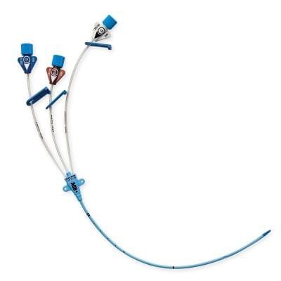
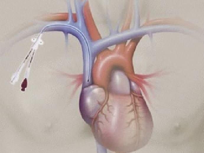
Now you can better utilize any central line for continuous ultrasound-based monitoring. Jugular, subclavian or even a peripherally inserted catheter can be used.
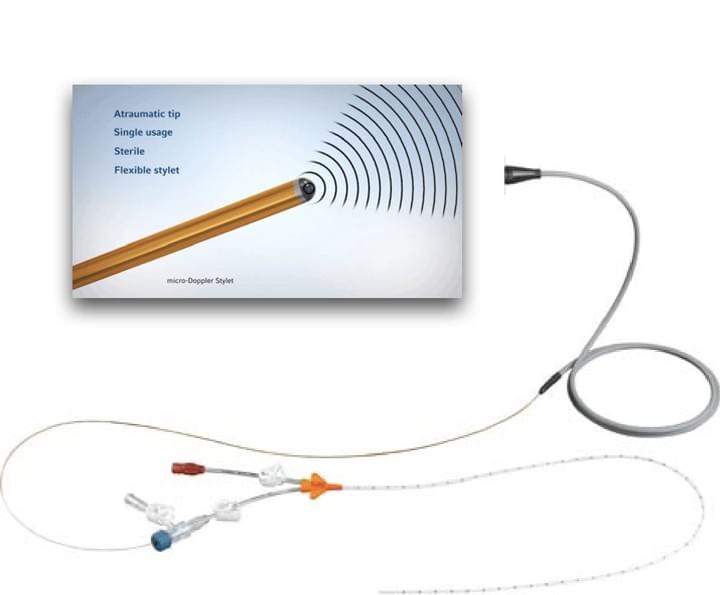
Miniature Doppler probe can be inserted via the distal lumen and easily positioned directly into the venous blood flow.

Compare the left side picture with the one on the right side
Getting both left-sided and right-sided perspective helps to discern better false positivity or false negativity of other monitoring methods used in hemodynamic therapeutical management.
Monitoring Superior Vena Cava Blood Flow Patterns
Using miniature Doppler probe insertable via any regular central line

Fluid Responsiveness
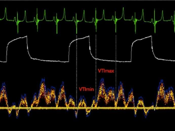
Well known and frequently used principle of respiratory flow variation originates in the thoracic section of the large veins. The Nilus algorithm calculates the flow variation right where it is detectable - in the Superior Vena Cava.
Veins are not able to adapt to blood loss as quickly as arteries do. The venous blood flow pattern is highly sensitive to changes caused by bleeding, vascular resistance, or right heart pressures.
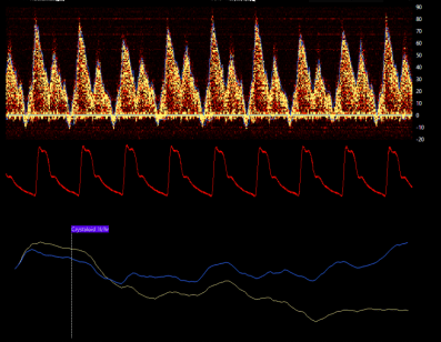
The picture shows a typical flow decrease (VTI min) during inspiration in the hypovolemic patient ventilated patient.
Alternatively, SVC blood flow velocity is a powerful surrogate to Stroke Volume during the fluid challenge.

Right Heart Performance
Echocardiography uses the Systolic/Diastolic wave ratios in ventricular performance assessment. The Nilus algorithm calculates the S/D ratio continuously and in real-time, providing a valuable parameter for the Right Heart performance.
The picture shows a healthy right heart pattern. During ventricular overload or failure, the pattern quickly changes. Most frequently, the S wave typically becomes less pronounced than the D wave.
Continuous monitoring adds significant value to the scheduled regular ultrasound examination, typically performed maximally a few times a day.

No added invasivness
Most of the hemodynamically unstable patients in need of continuous monitoring are having the central or PICC line in place. This line can now be even more utilized when a miniature Doppler probe is inserted into its distal lumen.
Insertion is hassle-free, and the optimal signal is stable and easy to obtain.
The probe can be cleanly secured to the catheter, easily disconnected or removed when needed.
- News & UpdatesSVC flow velocity monitoring can sensitively guide volume therapy during surgery Body is capable to maintain arterial pressure even during substantial hemorrhagic events. Latter sudden pressure drop to pre-shock or even shock happens when vasoconstriction can no longer maintain the desired...2018年9月28日Our pilot animal trial publication was done to prove the concept and collect key data for the algorithm development. This trial also triggered in-house development of a novel intravascular Doppler module demonstrating greater signal clarity and stability. Superior Vena Cava new fluid...Slide deck from EuroPCR 2015 Feasibility human data together with animal trial outcomes were presented by Dr. Kovarnik at EuroPCR in Paris. Human feasibility trial proved that clear and stable Doppler signal can be easily obtained and used by the device, when the probe is inserted via a...
Novel use of well-known technology
Designed for clinical and scientific use
Doppler Probe
Disposable 12MHz, 19G and smaller
ECG, AP Input
Directly from patient or analog signal slaving
Respiration
algorithmic or directly from the patient
Easy to Implement Into Your Current Workflow
Designed to fit any monitoring setup

Disposable Doppler Probe
Fast to Set Up
Miniature Doppler Probe reads the blood flow at the tip of central venous catheter

Modular Design
High Versatility
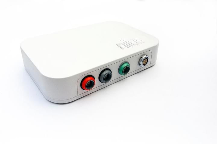
Nilus Databox contains Doppler module along with ECG, Pressure and Analog Input modules

Algorithmic processing
Simple Parameters
Fluid Responsiveness Index, Cardiac Output Surrogate, Right Heart Performance Index, Preload Parameters.

Ultrasound Made Monitoring Ready
Hands Free
Some of the well known ultrasound parameters can be now handily acquired continuously via stable central line probe.
- Contact Us
Learn more about Nilus Hemodynamic Monitor
© 2018


